Skip to content
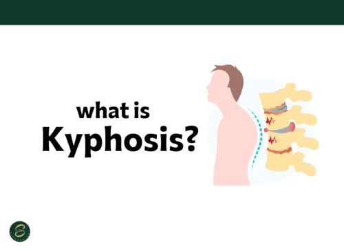
Kyphosis diagnosis is essential for detecting spinal deformities early and preventing long-term complications. Kyphosis is a forward rounding of the upper spine, often resulting in a noticeable hunchback appearance. It can develop due to poor posture, spinal abnormalities, osteoporosis, or congenital issues.
Common Symptoms of Kyphosis
 Diagnosing kyphosis starts with identifying key physical symptoms, which may include:
- Rounded upper back or hunched posture
- Back pain or stiffness
- Muscle fatigue in the shoulders and upper spine
- Reduced mobility or range of motion
- Uneven shoulder height
Medical Tests for Diagnosing Kyphosis
Healthcare professionals use several tests and imaging techniques to confirm kyphosis, such as:- Physical Examination: Observing spine curvature, posture, and mobility.
- X-rays: To measure spinal curvature (Cobb angle).
- MRI or CT scans: For more detailed views, especially if tumors or spinal cord issues are suspected.
- Bone Density Tests: Especially in older adults to detect osteoporosis-related kyphosis.
Types of Kyphosis Diagnosed
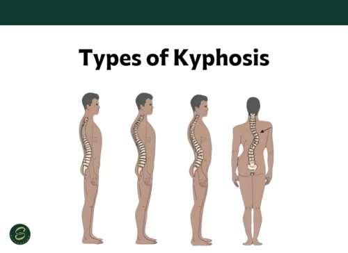 There are several distinct types of kyphosis, including:
- Postural Kyphosis: Often found in teenagers with poor posture; typically flexible and correctable.
- Scheuermann’s Kyphosis: A structural condition seen in adolescents.
- Congenital Kyphosis: Present at birth due to spinal malformation.
- Osteoporotic Kyphosis: Common in older adults due to weakened vertebrae.
Corrective Solutions for Kyphosis
Once diagnosed, kyphosis treatment depends on the severity and underlying cause. Common corrective strategies include:1. Physical Therapy & Exercise
Strengthening the back muscles and improving flexibility can significantly reduce spinal curvature and pain.2. Posture Correction Tools
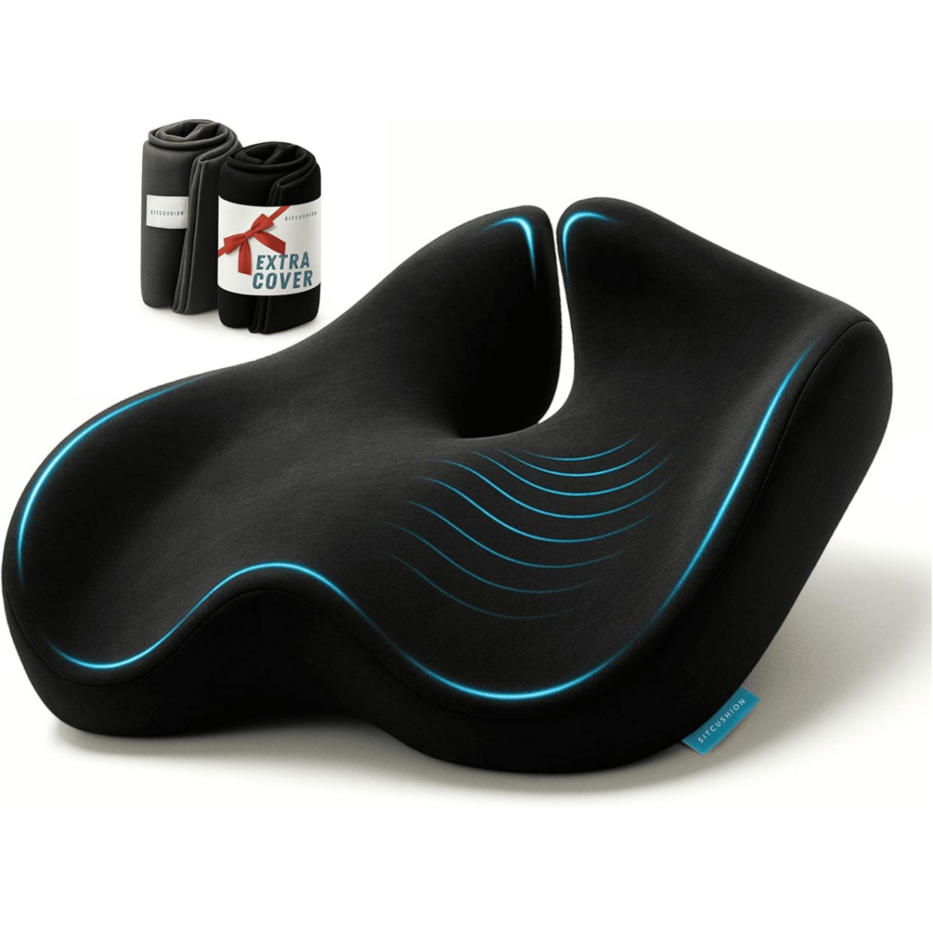
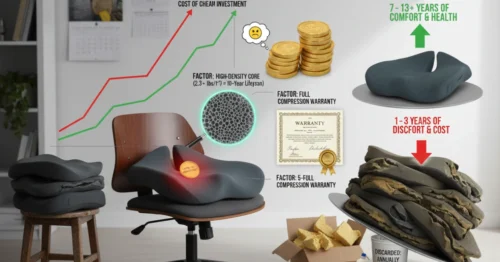
Ergonomic tools like seat cushions and back support devices can improve posture during daily sitting, especially for those with sedentary lifestyles. Check out our ergonomic seat cushions designed to promote spinal alignment and reduce pressure.3. Bracing
For growing children and teens, a spinal brace may help prevent further curvature.4. Medication or Supplements
Especially in osteoporotic cases, calcium, vitamin D, or bone-strengthening medication may be recommended.5. Surgical Options
In severe cases, spinal fusion surgery may be required, especially when there’s nerve compression or progressive deformity.Kyphosis and Everyday Comfort
Even mild kyphosis can lead to discomfort when sitting for extended periods. That’s where supportive cushions come in. Using a well-designed cushion helps:- Align your spine naturally
- Reduce pressure on the tailbone
- Support long hours of sitting whether at work, in the car, or at home